42 brain mri with labels
Brain lobes - annotated MRI | Radiology Case | Radiopaedia.org BSCI-14. SYNTHETIC METASTATIC BRAIN DISEASE MRI IMAGES CREATED USING A GENERATIVE ADVERSARY NETWORK TO OVERCOME DEEP MACHINE LEARNING CHALLENGES IN HEALTHCARE. Dai et al., Neuro-Oncology Advances, 2019. Atlas of BRAIN MRI - W-Radiology Brain magnetic resonance imaging (MRI) is a common medical imaging method that allows clinicians to examine the brain's anatomy (1). It uses a magnetic field and radio waves to produce detailed images of the brain and the brainstem to detect various conditions (2).
Frontiers | 101 Labeled Brain Images and a Consistent Human Cortical ... Labeled anatomical subdivisions of the brain enable one to quantify and report brain imaging data within brain regions, which is routinely done for functional, diffusion, and structural magnetic resonance images (f/d/MRI) and positron emission tomography data.
Brain mri with labels
Cross-sectional anatomy of the brain - e-Anatomy - IMAIOS Axial MRI Atlas of the Brain. Free online atlas with a comprehensive series of T1, contrast-enhanced T1, T2, T2*, FLAIR, Diffusion -weighted axial images from a normal humain brain. Scroll through the images with detailed labeling using our interactive interface. Perfect for clinicians, radiologists and residents reading brain MRI studies. Brain MRI segmentation | Kaggle Journal of Neuro-Oncology, 2017. This dataset contains brain MR images together with manual FLAIR abnormality segmentation masks. The images were obtained from The Cancer Imaging Archive (TCIA). They correspond to 110 patients included in The Cancer Genome Atlas (TCGA) lower-grade glioma collection with at least fluid-attenuated inversion ... Brain MRI Dataset | Kaggle Brain MRI Dataset | Kaggle. Haşim Mumcu · Updated 3 years ago. arrow_drop_up. 5. New Notebook. file_download Download (8 MB)
Brain mri with labels. 101 Labeled Brain Images and a Consistent Human Cortical Labeling ... Labeled anatomical subdivisions of the brain enable one to quantify and report brain imaging data within brain regions, which is routinely done for functional, diffusion, and structural magnetic resonance images (f/d/MRI) and positron emission tomography data. Head MRI: Purpose, Preparation, and Procedure - Healthline Magnetic resonance imaging (MRI) of the head is a painless, noninvasive test that produces detailed images of your brain and brain stem. An MRI machine creates the images using a magnetic field and... Researchers automate brain MRI image labeling, more than 100,000 exams ... Researchers have automated brain MRI image labeling, needed to teach machine learning image recognition models, by deriving important labels from radiology reports and accurately assigning them to... Brain MRI Atlas on the App Store Brain MRI Atlas is a FREE app that allows you to navigate through hundreds of of labeled brain structures. It is designed for all healthcare professionals as an interactive study and reference tool. Program Features: - Serial sequential axial T2 FLAIR images of the brain. - Structure labels organized by category.
Automated MRI image labelling processes 100,000 brain exams in under 30 ... Researchers from the School of Biomedical Engineering & Imaging Sciences at King's College London have automated brain MRI image labeling, needed to teach machine learning image recognition models,... MR Image Classification for Brain Tumor Texture Based on Pseudo-Label ... MR Image Classification for Brain Tumor Texture Based on Pseudo-Label Learning and Optimized Feature Extraction Comput Math Methods Med. 2022 Apr 4;2022:7746991. doi: 10.1155/2022/7746991. ... First, for the small sample of pituitary tumor MRI image data, the T1 and T2 sequence data are uneven or missing; we used the CycleGAN model to perform ... Brain: Atlas of human anatomy with MRI - e-Anatomy - IMAIOS MRI Atlas of the Brain. This page presents a comprehensive series of labeled axial, sagittal and coronal images from a normal human brain magnetic resonance imaging exam. This MRI brain cross-sectional anatomy tool serves as a reference atlas to guide radiologists and researchers in the accurate identification of the brain structures. Deep learning to automate the labelling of head MRI datasets for ... manually labelling mri scans appears to be particularly laborious due to (1) the superior soft-tissue contrast of mri which enables more refined diagnoses compared with other imaging modalities such as computed tomography; and (2) the use of multiple, complementary imaging sequences so that a larger number of images must be scrutinised per …
UCLA Brain Mapping Center - ICBM Template To view both the structural MRI and the labels launch the program typing Display icbm_template.mnc -label icbm_labels_corrected.mnc. The opacity of the labels can be set in the Colour Coding menu. The number of each label appears at the bottom left of the orthogonal views window. What Does a Brain MRI Show? - San Diego Health What does a brain MRI show? The answer is, unfortunately, not very. MRI scans (magnetic resonance imaging) have been around for decades, and the technology has been steadily improving. Today, a brain MRI test can identify whether or not a person has a stroke, or if the person has suffered a traumatic brain injury, or if the person is suffering ... The MRI Dataset with labels. | Download Scientific Diagram Download scientific diagram | The MRI Dataset with labels. from publication: Detection of brain abnormality by a novel Lu-Net deep neural CNN model from MR images | The identification and ... NITRC: Manually Labeled MRI Brain Scan Database: Tool/Resource Info This is a continuously growing and improving database of high-quality neuroanatomically labeled MRI brain scans, created not by an algorithm, but by neuroanatomical experts. All results are checked and corrected. Regions of interest include the usual sub-cortical structures (thalamus, caudate, putamen, hippocampus, etc), along with ventricles ...
Opensided MRI - Shields
CaseStacks.com - MRI Brain Anatomy Labeled scrollable brain MRI covering anatomy with a level of detail appropriate for medical students. Show/Hide Labels. MRI Brain Anatomy. Back to Anatomy Overview. ... Labelled radiographs and CT/MRI series teaching anatomy with a level of detail appropriate for medical students and junior residents. Pelvis. Pelvic MRI anatomy

Dr Balaji Anvekar FRCR: Miliary Tuberculosis of CNS
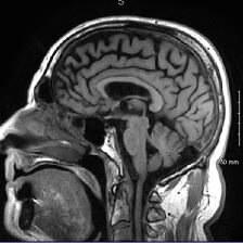
Brain MRI: How to read MRI brain scan | Kenhub MRI is the most sensitive imaging method when it comes to examining the structure of the brain and spinal cord. It works by exciting the tissue hydrogen protons, which in turn emit electromagnetic signals back to the MRI machine. The MRI machine detects their intensity and translates it into a gray-scale MRI image.

Marks in the Margin: An MRI
Labeled imaging anatomy cases | Radiology Reference Article ... This article lists a series of labeled imaging anatomy cases by body region and modality. Brain CT head: non-contrast axial CT head: non-contrast coronal CT head: non-contrast sagittal CT head: angiogram axial CT head: angiogram coronal CT...

Advanced MRI brain scans - Stock Image - P332/0521 - Science Photo Library
Arterial spin labeling MRI: Clinical applications in the brain THE BRAIN is a unique organ, housed in a rigid calvarium and dependent on high rates of blood flow (average 50 mL/100 g/min). ... (MRI) method that uses magnetically labeled blood water as a flow tracer, providing CBF images of the brain. Moreover, if certain conditions are met it can potentially also provide an absolute, quantifiable CBF ...

MRI of the Human Brain - College of Sciences
Labeled MRI Brain Scans - Neuromorphometrics We can also label scans that you provide and we are very interested in labeling white matter anatomy as seen in diffusion-weighted MRI scans. If you want an aggregate version of our data, we can provide it as a probabilistic atlas. The cost to label a single scan is $2449 (USD).

Dr Balaji Anvekar's Neuroradiology Cases: Focal Cortical Dysplasia MRI
brain anatomy | MRI coronal brain anatomy | free MRI cross sectional ... This MRI brain coronal cross sectional anatomy tool is absolutely free to use. Use the mouse scroll wheel to move the images up and down alternatively use the tiny arrows (>>) on both side of the image to move the images.>>) on both side of the image to move the images.

AdvantageImaging
GitHub - yunshiuan/label4MRI: Label the brain MNI coordinate by AAL/BA ... MRI-labeling: label human brain MRI image by AAL/BA system Under the R program environment,input an MNI coordinate, output the corresponding AAL(Automated Anatomical Labeling) and BA (Brodmann area) brain region name. More importantly, if the coordinate does not match a brain region defined by AAL/BA (e.g., white matter), the package help find the closest brain region with the corresponding ...

22C+: Small Things and the Big Picture
AutoComBat: a generic method for harmonizing MRI-based radiomic ... Furthermore, we have developed AutoComBat, which aims to automatically determine the batch labels, using either MRI metadata or quality metrics as inputs of the proposed constrained clustering. A ...

066 The paradoxical response: the development of intracranial tuberculomas during treatment for ...
MRI anatomy | free MRI axial brain anatomy MRI anatomy | free MRI axial brain anatomy This MRI brain cross sectional anatomy tool is absolutely free to use. Use the mouse scroll wheel to move the images up and down alternatively use the tiny arrows (>>) on both side of the image to move the images.

Brain At 22 Years | Volume Rendering of an MRI scan of the b… | Flickr
101 labeled brain images and a consistent human cortical labeling ... given how difficult it is to label brains, the mindboggle-101 dataset is intended to serve as brain atlases for use in labeling other brains, as a normative dataset to establish morphometric variation in a healthy population for comparison against clinical populations, and contribute to the development, training, testing, and evaluation of …

Cross-sectional anatomy of the brain | Brain, Mri, Interventional radiology
Brain MRI Dataset | Kaggle Brain MRI Dataset | Kaggle. Haşim Mumcu · Updated 3 years ago. arrow_drop_up. 5. New Notebook. file_download Download (8 MB)

22C+: Small Things and the Big Picture
Brain MRI segmentation | Kaggle Journal of Neuro-Oncology, 2017. This dataset contains brain MR images together with manual FLAIR abnormality segmentation masks. The images were obtained from The Cancer Imaging Archive (TCIA). They correspond to 110 patients included in The Cancer Genome Atlas (TCGA) lower-grade glioma collection with at least fluid-attenuated inversion ...

Applying machine learning for clinical decision support - Vector blog
Cross-sectional anatomy of the brain - e-Anatomy - IMAIOS Axial MRI Atlas of the Brain. Free online atlas with a comprehensive series of T1, contrast-enhanced T1, T2, T2*, FLAIR, Diffusion -weighted axial images from a normal humain brain. Scroll through the images with detailed labeling using our interactive interface. Perfect for clinicians, radiologists and residents reading brain MRI studies.

Chiari 1 Malformation | I'm trying to figure out how to add … | Flickr

Spinal nerve levels, sagittal MRI - Stock Image C030/3581 - Science Photo Library
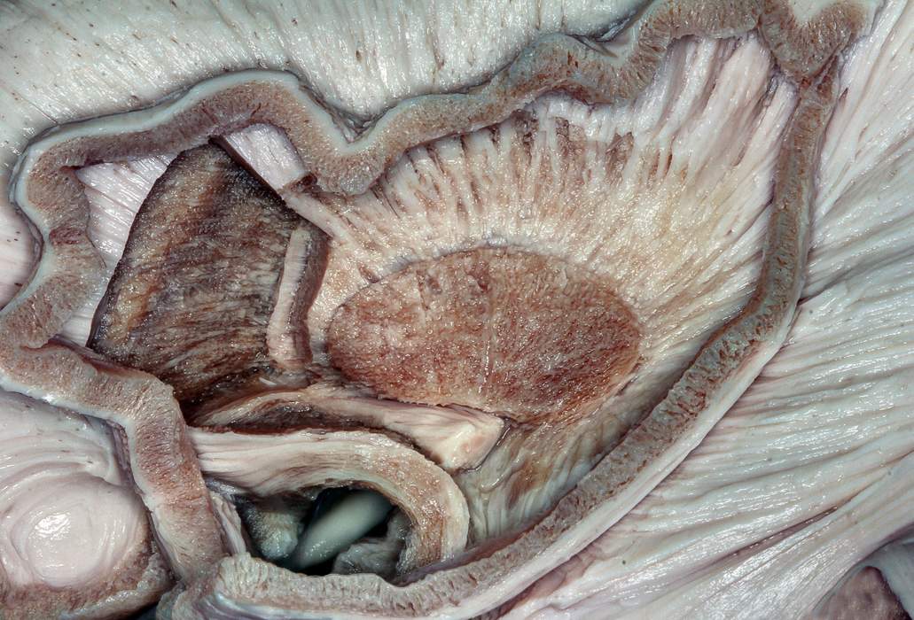
Sagittal View of the Basal Ganglia and Internal Capsule | Neuroanatomy | The Neurosurgical Atlas ...

Dr Balaji Anvekar FRCR: Joubert syndrome MRI
Post a Comment for "42 brain mri with labels"